Our ophthalmologist can diagnose common retinal disorders such as macular degeneration and diabetic eye disease. Along with careful retinal examination, the Spectral domain Ocular Coherence Tomography unit (SD-OCT) by Heidlberg Engineering can detect and monitor for retinal conditions.
Macular Degeneration
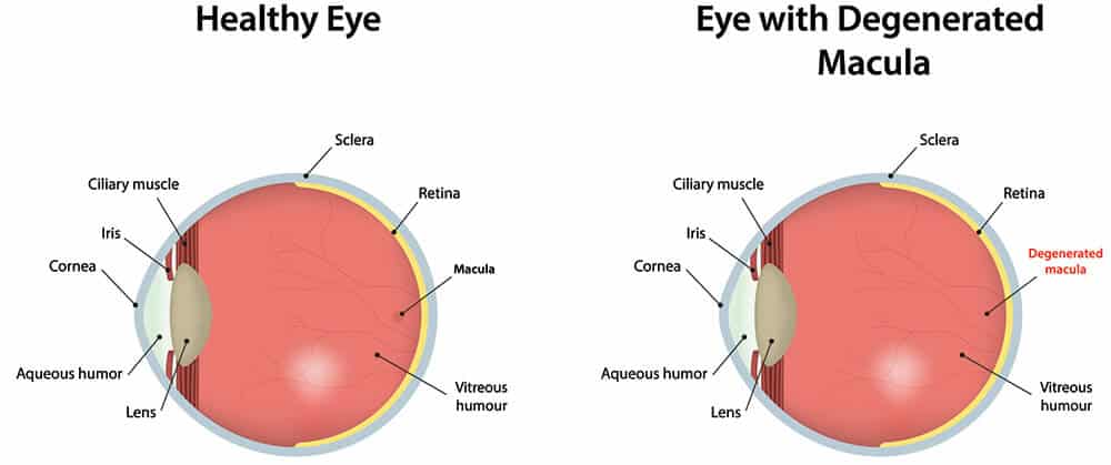
Age related macular degeneration (ARMD) can have detrimental effects on vision. It is a breakdown on the nutrient layer of the retina in the back of the eye. Family history, sun exposure, caucasian descent, smoking history, and cardiovascular disease are all risk factors. Most often ARMD is of the dry variety and requires only periodic monitoring. Less commonly it can develop into the “wet” or neovascular variety which implies the bleeding or swelling is occurring within the retina. The SD-OCT can pick up subtle changes not observable on examination alone and frequent retina evaluations can help ensure early changes are detected and that vision is preserved.
The Age-Related Eye Disease Study (AREDS) showed that eye-specific vitamins can slow down the progression of ARMD for those with moderate to advanced disease. It is not known, however, if the use of the vitamins can prevent ARMD in those without the condition. More recently, the AREDS-2 study is showing the addition of Lutein and Zeazanthine are also helpful in slowing the process down.
Diabetic Retinopathy
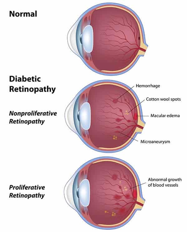
Diabetes can cause Retinopathy- a microscopic leakage of the capillaries within the retina, which can lead to vision threatening side effects. The American Medical Academy recommends at least yearly screening for this common and often symptomless condition. Diabetes can also lead to Macular Edema (CME). Yearly examination in combination with the SD-OCT can detect early changes which may lead to vision compromise. Since treating clients throughout the Syracuse, Oneida and Liverpool NY areas, Dr. Cecchi will ensure to communicate with your internist or general practitioner to ensure that your file is complete with your yearly eye exam.
What are the stages of Diabetic Retinopathy?
There are four stages of diabetic retinopathy to be aware of:
Mild nonproliferative diabetic retinopathy
Moderate nonproliferative diabetic retinopathy
Severe nonproliferative diabetic retinopathy
Proliferative diabetic retinopathy
Nonproliferative diabetic retinopathy is an early version of the disease, whereas proliferative is advanced. The best way to find out what stage you are in is to get specialized eye tests to help your eye doctor determine what stage your diabetic retinopathy is at.
What are the Symptoms of Diabetic Retinopathy?
If you’re experiencing these symptoms, you may be developing diabetic retinopathy.
Spots or floaters
Blurred vision
Fluctuating vision
Impaired color vision
Dark or empty areas in your vision
Vision loss
However, suppose you are not experiencing these symptoms. In that case, you may still be at risk for diabetic retinopathy as it does not have symptoms in its early stages of development.
How do you treat diabetic retinopathy?
Laser surgery treatment options are available to stop or slow the progression of this disease. There are also surgical procedures and medicine injections that can be performed. These solutions may not cure the condition, although it may slow or stop its progression.
One of the most significant ways to treat diabetic retinopathy is to take steps to prevent it.
Even if you are not experiencing symptoms, you may still be at risk. Some ways to prevent diabetic retinopathy include managing your diabetes and/or blood sugar level, lowering your cholesterol, stop smoking tobacco, and monitor any vision changes.
What causes diabetic retinopathy?
Diabetic retinopathy is caused by blood vessels in the light-sensitive tissue back of the eye or the retina.
Those who have type 1 or type 2 diabetes are at risk for developing this condition. The longer you have it and the higher your blood sugar is, the more likely you are to develop diabetic retinopathy.
Floaters
Floaters develop within the vitreous cavity of the eye. The central part of the eye is filled with a clear, jelly like substance known as the vitreous humor. Over time, collagen strands within the gel starts to shrink, forming wisps and strands which appear to float in front of the vision. If the gel shrinks completely, it pulls away from its internal attachments to the retina, and may appear as a denser opacity that may drift in front of the vision. Dr. Cecchi can diagnose if this is occuring.
Retinal Tear/Retinal Detachment
Retinal tear can occur when the vitreous gel, when shrinking, causes traction on the retina, the delicate neurosensory layer in the back of the eye. Retinal tear is often associated with flashes and a sudden increase in floaters. If diagnosed, immediate referral to a retina specialist is important to treat this potentially vision threatening condition.
If the retinal tear is more advanced, a retina detachment often follows. A detached retina is a serious condition which usually leads to permanent vision loss if left untreated.